
Henry Quach
Optical EngineerSurface Metrology of a Butterfly's Wing
Introduction
You can buy bugs on Etsy. As it turns out, the most popular insect seller on Etsy is a pair of entomologists based in Sahuarita, just 15 miles south of the university. Naturally, my labmate Hyukmo and I bought butterflies from them so we could examine the scale structures under two microscopes at opposite ends of the price spectrum. In this short blog post, I'll show you what we saw.
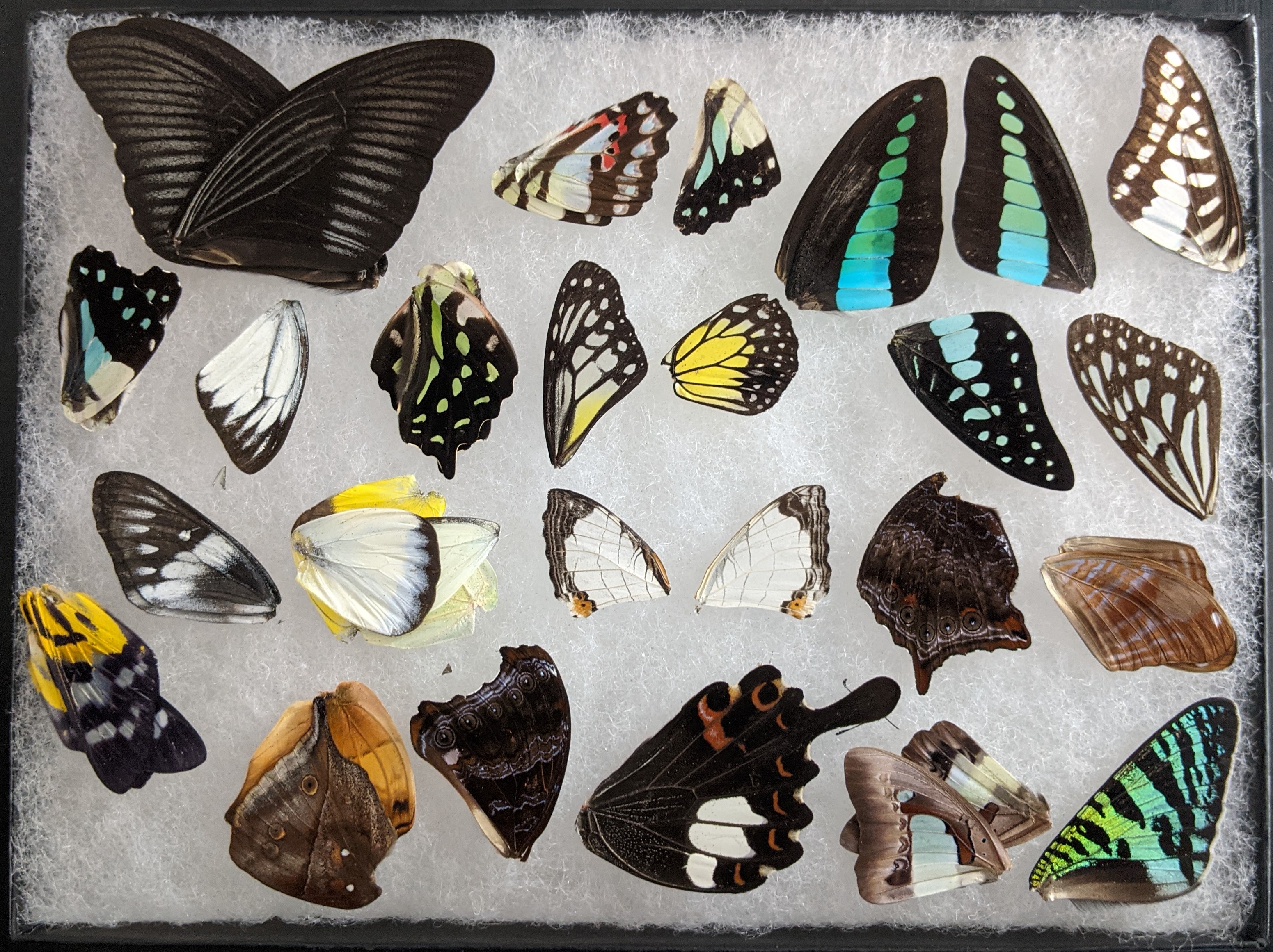
Figure 1. I bought BicBug's 'Lot of 50 Butterfly and Moth Wings', kind of like a booster pack for Magic TCG but much cooler. I ended up with a variety of wings, including that of a swallowtail, monarch, monarch mimic, autumn leaf, straight line map wing, and sunset moth.
Digital Microscope - Well, a Macro Lens
If you search on Amazon for 'USB Microscope', you will find limitless variety of rebranded Chinese 'bullet-style' microscopes between $20 and $50. They make claims for magnifications between 10x and 4000x. Of course, these claims are baseless as magnification is easy, but resolving power is hard. The 'microscope' is simply a macro lens that slides along a helical cam mechanism. This allows you to change conjugate object planes to the CMOS detector. Still, for the price, the ability to see tiny details can't be beat. These are highly common in home electronics labs for soldering stations.
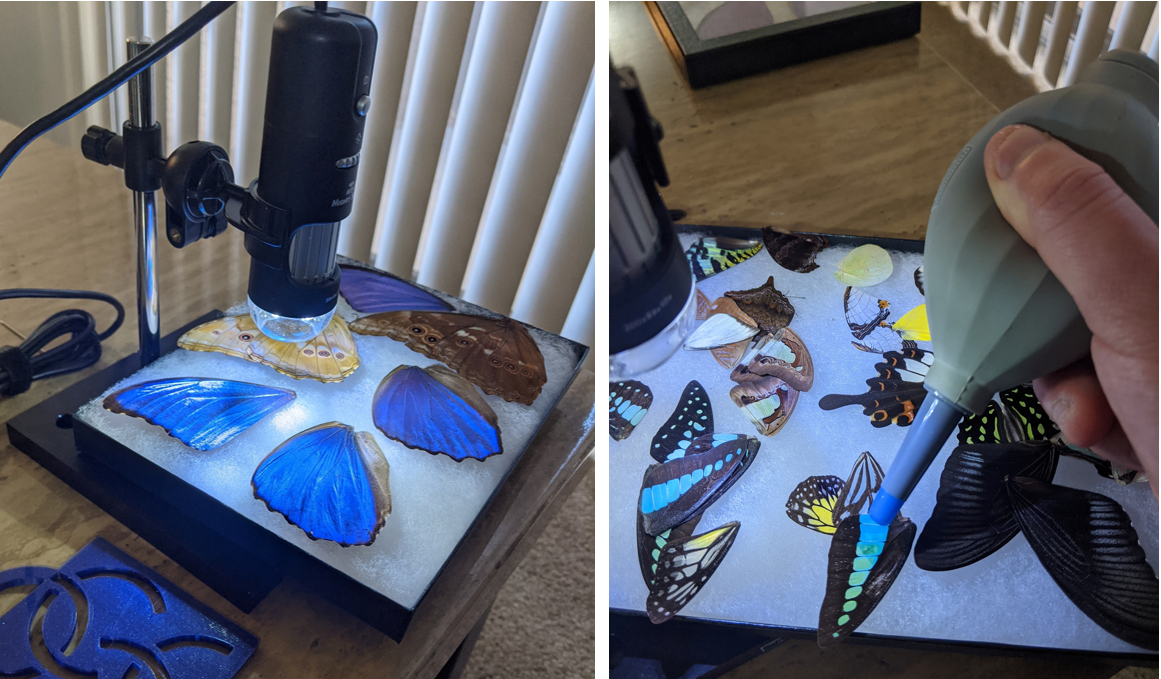
Figure 2. My bullet microscope was manufactured by Aven Tools, which is known for their high quality tweezers. This is the microscope stocked by McMaster-Carr, which by the way, does not typically label their parts with brand names unless necessary. I was expecting the Dino-Lite, which is among the most reputable bullet-style microscopes. Before examining any insect parts beneath the microscope, I used a dust blower as to remove any surfacial dust that might possibly get onto the objective lens. THe two figures show my Blue Morphos (Morpho didius; Morpho godarti asarpai, both from Peru) as well as my framed collection of miscellaneous wings.
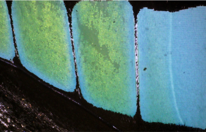
Figure 3. The wings of the Graphium antheus are green and blue. The size of each scale is on the order of 30-80 microns. Butterfly wing scales are made of chitin and do not regenerate over the lifetime of the butterfly. If you were to touch a butterfly's wings with your oily fingers, some scales could rub off :(.
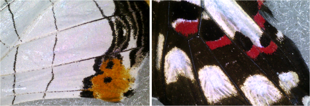
Figure 4. The scales of the Straight Line Map Wing butterfly (now one of my favorite butterflies because of its beautiful geometric patterning) and the 'furry'-looking wings of the imitation monarch.
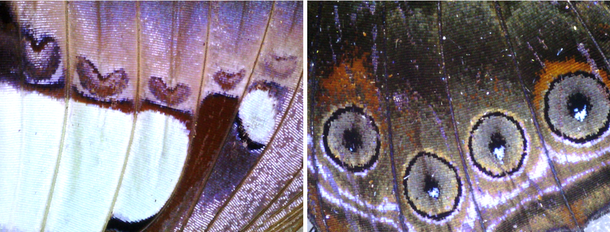
Figure 5. Observe how each scale is like a 'pixel' and that the borders of the hearts and circles can be just one pixel wide. It is stunning that these precise visual patterns can at all be encoded into the genetic material of a living creature.
White Light Microscope
An interference microscope leverages the temporal coherence of a white light source to reconstruct a surface - specular or diffuse. These vertical scanning interferometers find an irradiance signal through focus. At a single pixel, we see many wavelengths obtaining a single precise 'coherence envelope', which is identifiable with maximum contrast for a specific single height plane. Each wavelength creates its own interference fringe pattern but equal optical path among all wavelengths is only achieved when the sample is at a specific height. Some advantages are that there are no spurious fringes or speckle, which is present in typical quasimonochromatic interferometers. However, some disadvantages are that they are limited by the resolution of the height step size and only so much area can be scanned at a time.
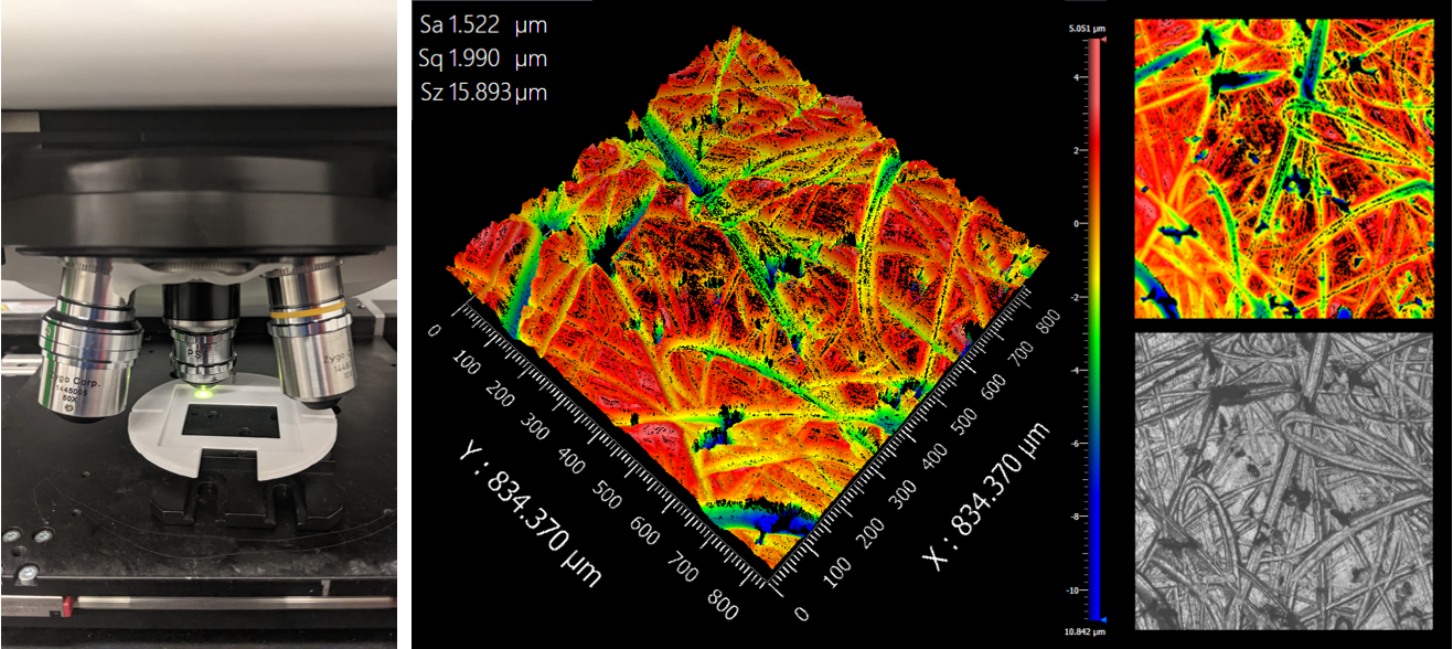
Figure 6. I used the Zygo Newview 8300 for the next part. Note that I obliterated all dust from the butterflies before even approaching the machine as to mitigate all risk of damaging the objectives. The measurements on the right show the surface profile of anodized black aluminum, NOT 3D printed plastic.
The Morpho Butterfly
Morpho, morpho, morpho. Morpho.
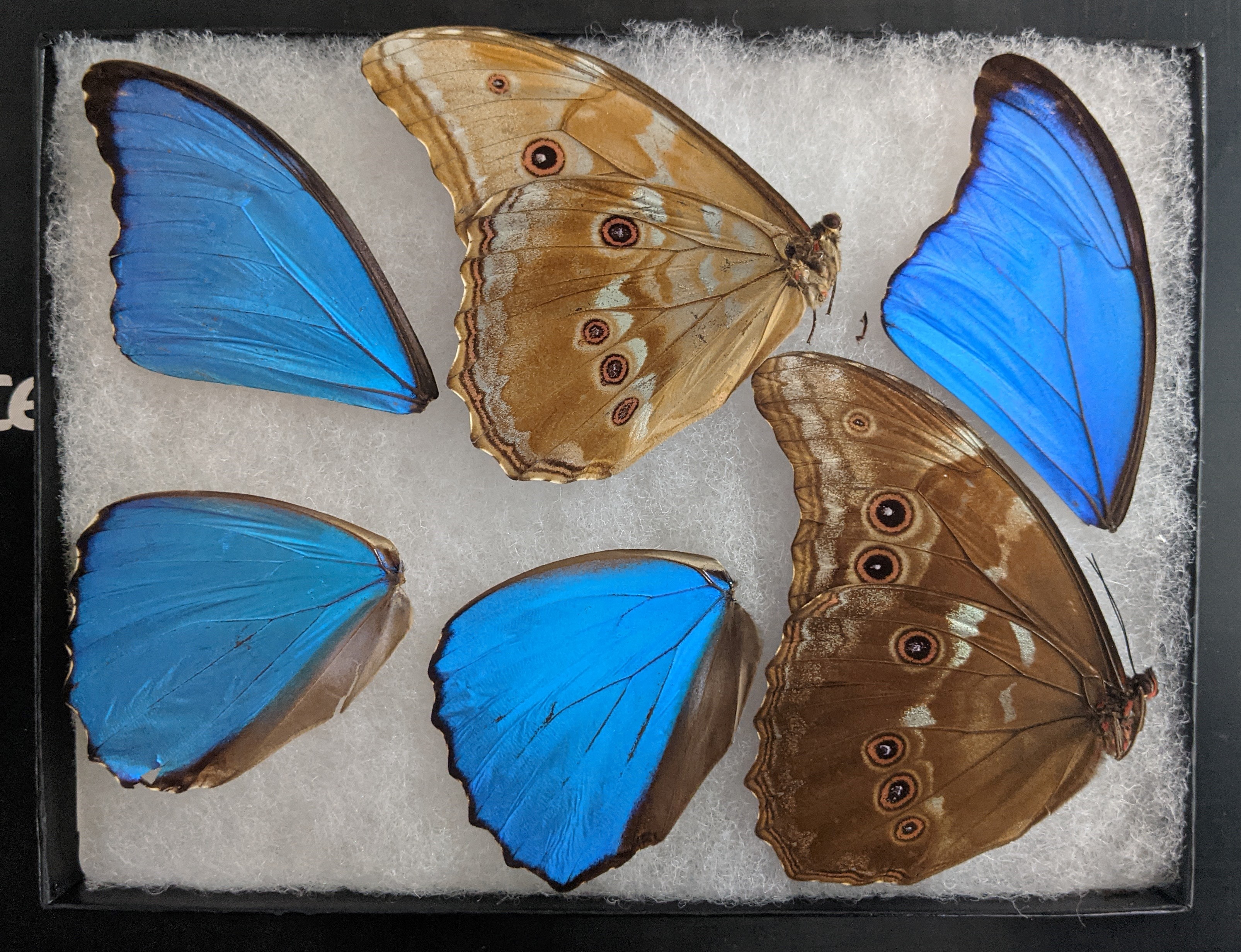
Figure 7. The blue from the Blue Morpho is not from pigment, but diffractive volumetric structures within each scale. If you step back and look from oblique angles, the butterfly turns violet.
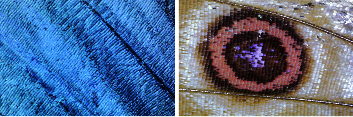
Figure 8. Under the digital USB microscope - both sides of the wings. Predators such as birds have a harder time distinguishing blue wings from the rest of the sky at higher altitudes. From below, the dark annuli look like eyes.
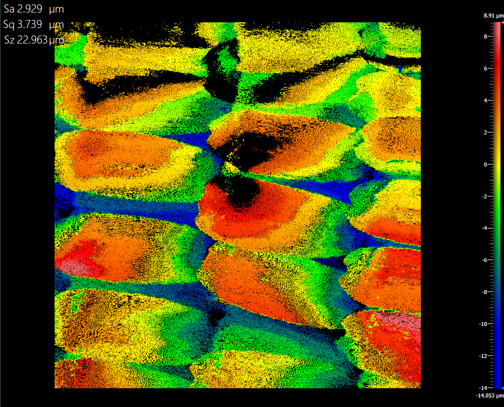
Figure 9. This patch of information is approximately 834 um by 834 um. The surface is pretty rough - up to ~ 4 microns RMS. Black patches were out of range of the scanning interferometer. The Mirau objective was used to obtain this reconstruction.
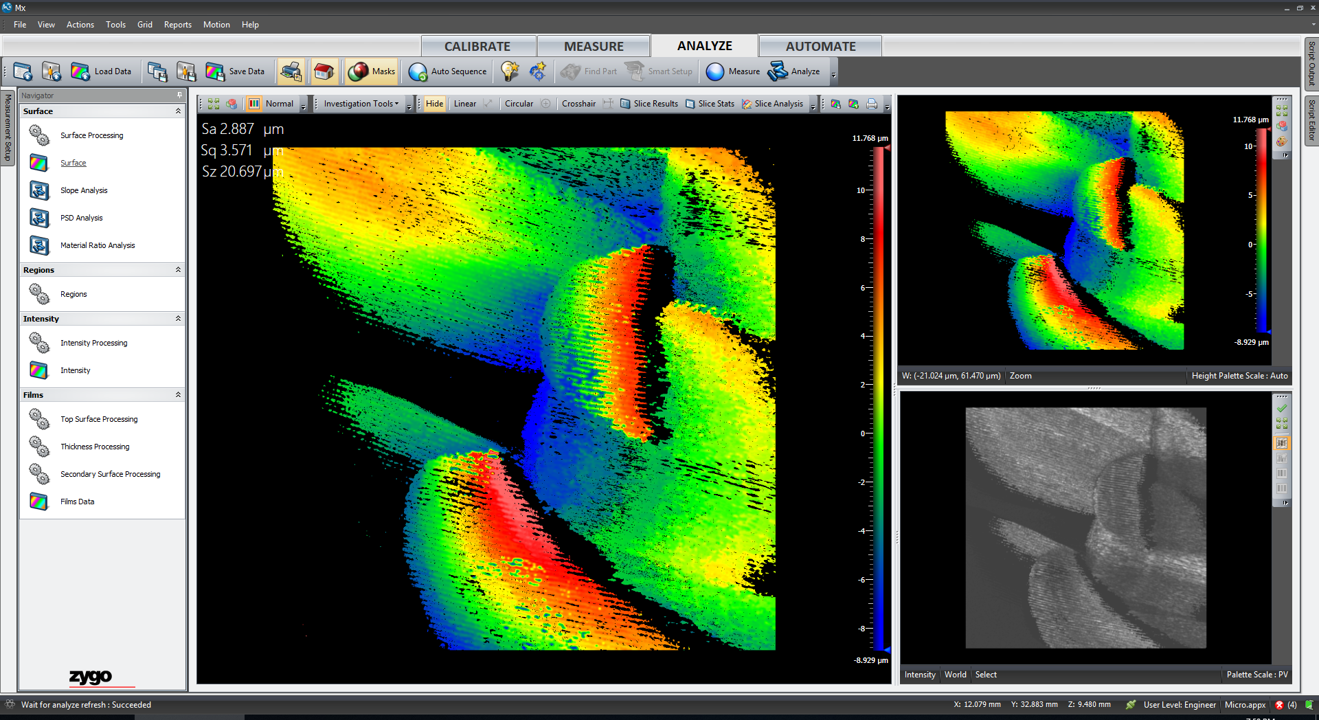
Figure 10. A slice was drawn across the tiny micro-ridges along one scale. It is because of this sinusoidal ridge feature that the butterfly scale isn't so specular.
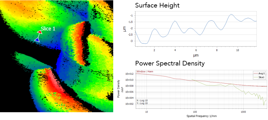
Figure 11. Spacing along a ridge is on the order of a micron. Fascinating. The power spectral density is flatter than one might expect.
Nabakov's Butterfly
I specifically obtained the Papilio machaon because it is one of the butterflies mentioned by Nabakov in his works. Nabakov was a well-known amateur lepidopterologist, or entomologist who studies butterflies. This specimen was obtained in Pima County, AZ in Sahuarita. This specimen is also known as Baird's Old World Swallowtail.
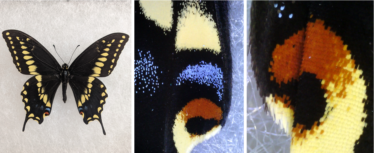
Figure 12. Look at the beautifully resolved scale structures - yellow, orange, and blue. They pop out more at you because they are sloped upwards.
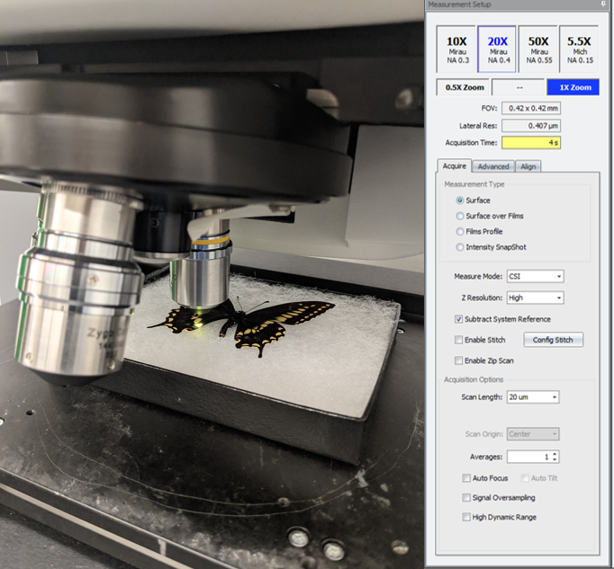
Figure 13. Examining this under the 20x Mirau objective, safely, of course.
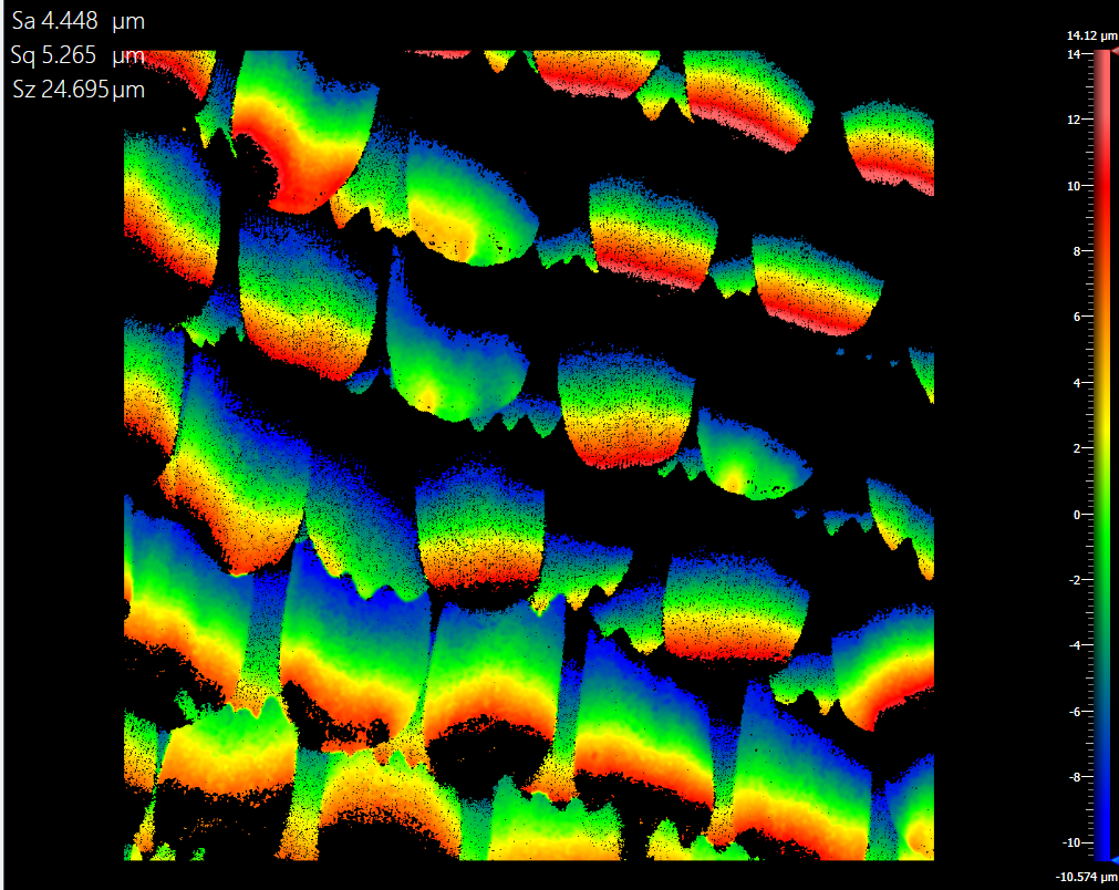
Figure 14. One of the challenges for the specimen, in comparison with the Blue morpho reconstructions, was that the scales were so strongly sloped upwards. This increased the dynamic range of heights - one that cannot be easily minimized without significantly tilting the entire specimen. Black regions are cloaks of NaNs because the sloped scales cast shadows and vignette all possibility of white light interference.
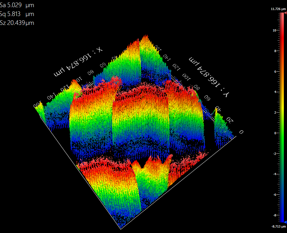
Figure 15. Under the 50X Mirau objective, the fine undulating patterns of each scale is visible. The 3D visualization makes each scale look like a Ruffle. Now the FOV only covers a 160 x 160 um square, but the non-uniform spacing of scales is more clearly evident.
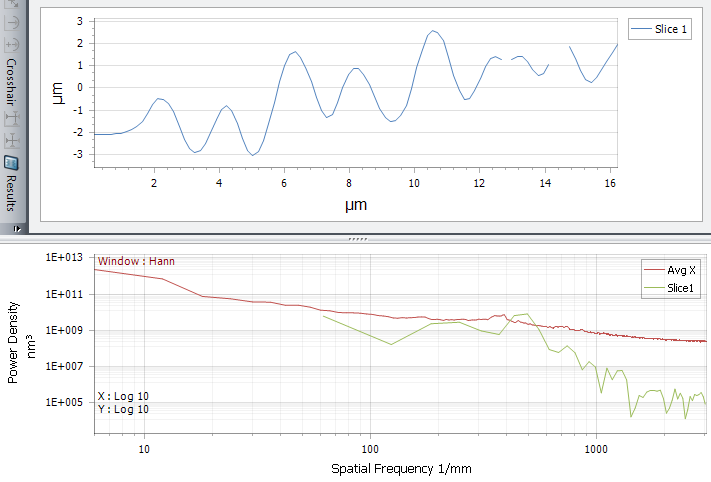
Figure 16. The first figure shows the similar 1 micron undulation seen in the Blue Morpho's Scale. The power spectral density is similarly 'flat'.
Conclusions
Anyway, this was a fun project! I hope to add to this with the teardown of a cheap USB bullet microscope, as well as spatial frequency analysis of the wings. I saved all the datasets and will spatially analyze them with a software called SAGUARO developed by my lab.